Medical College of Wisconsin Ophthalmology and Visual Sciences Research Study - Retinitis Pigmentosa & Usher Syndrome
This research study is being conducted to examine retinal structure and function in subjects with retinitis pigmentosa and Usher syndrome.
What is retinitis pigmentosa & Usher syndrome?
Retinitis pigmentosa (RP) is a degenerative eye condition that can cause severe vision loss. RP is noted by pigment changes in the back of the eye and a degeneration of the cells needed for vision. Usher syndrome is similar to RP but is also associated with hearing loss.
What is involved in our research?
This non-invasive imaging study may include one or more visits to the Eye Institute. Research volunteers will be asked to complete an ocular health questionnaire and a series of vision tests, and may be asked to give a blood sample, saliva sample, or buccal swab from the inside of the cheek to test for the mutations that cause retinitis pigmentosa and Usher syndrome. In addition, we will use various imaging devices to take pictures of the eye – including optical coherence tomography (OCT), fundus photography, and adaptive optics retinal imaging. At least one eye will be dilated during the visit. There is no direct health benefit to volunteers. Each visit typically takes about 3 hours.
Research volunteers will receive $15 per hour for their time.
You may be eligible to participate in this study if you meet these criteria.
- You are at least 5 years old.
- A doctor has told you that you have retinitis pigmentosa or Usher syndrome.
- You are willing and able to provide informed consent.
- Your vision is better than 20/200 in at least one eye.
More Information:
Contact: aoip@mcw.edu (414) 955-AOIP
IRB Approval: PRO00017439
5/18/2013
Contact Ophthalmology
For patient care inquires, call us at (414) 955-2020 or use MyChart. Email is for research and education inquiries only.
Eye Institute Location
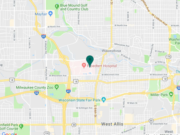
925 N. 87th St.
Milwaukee, WI 53226
Appointments
(414) 955-2020
(414) 955-6166 (fax)
Continuing Medical Education
Renee Ware
(414) 955-2086
Medical Education Coordinator
Associate Director of Development - Ophthalmology
Sarah Walker
Refer to Us - Consultation requests
Patient Referral Form (PDF)
Fax to (414) 955-0136
Emergent Requests
Within 48 hours call
(414) 955-2020
Research
Vesper Williams
(414) 955-7862
Advanced Ocular Imaging Program
(414) 955-2647
Eye Institute Executive Director (Administrator)
Shannon Dreier


