Ophthalmology_Hero Image 2

Case Study 10 - CC: Blurry vision in the left eye for 2 weeks

all
Patient Visit

Patient History

HPI:
A 72 year-old female retired CEO reports that approximately 2 weeks ago she noticed the onset of decreased vision in her left eye associated with a “black spot” close to the center of her vision. The spot is present with both near and far vision and does not move away. The vision has been slightly blurred for a while. She denies any recent trauma, eye pain, redness or discharge. Peripheral vision is normal. She has not had an eye exam in 5 years.

Past Ocular History:
No prior eye surgeries. No hx of eye trauma, amblyopia or strabismus.

Ocular Medications:
None

Past Medical History:
Hypertension, hypercholesterolemia, coronary artery disease s/p myocardial infarction s/p stenting 5 years prior.

Surgical History:
C-sections x 2, appendectomy 15 years ago

Past Family Ocular History:
No evidence of blinding diseases, glaucoma or macular degeneration

Social History:
Past 50 pack/year smoking history. No alcohol or drug use. Currently retired and living with her husband

Medications:
Aspirin, Plavix, Lipitor, Lisinopril, Metoprolol

Allergies:

ROS:
Denies recent illness or any new CNS, heart, lungs, GI, skin or joint symptoms.

Ocular Exam

Visual Acuity (cc):
OD: 20/30
OS: 20/100

IOP (tonoapplantation):
OD: 16 mmHg
OS: 17 mmHg

Pupils:
Equal, round and reactive to light, no APD OU

Extraocular Movements:
Full OU. No nystagmus.

Confrontational Visual Fields:
Full to finger counting OU.

External:
Normal, both sides

Slit Lamp:

Lids and Lashes Normal OU
Conjunctiva/Sclera Normal OU
Cornea Clear OU
Anterior Chamber Deep and quiet OU
Iris Normal OU
Lens 1+ NSC OU
Anterior Vitreous Clear OU

Dilated Fundus Examination:

OD Clear view, CDR 0.5 with sharp optic disc margins, flat macula, scattered large soft drusen within the arcades, normal vessels and peripheral retina
OS Clear view, CDR 0.6 with sharp optic disc margins, flat macula, scattered soft drusen within the arcades, 1 disc area of subretinal hemorrhage in macular area near fovea

Other:

Amsler grid:
OD: Normal
OS: Blurry spot near the center of the grid with wavy lines

Diagnosis and Discussion

Diagnosis
Wet age-related macular degeneration

Discussion

Differential Diagnosis:
The appearance of bilateral soft drusen and an area of subretinal hemorrhage in the left eye is consistent with the diagnosis of wet age-related macular degeneration. The subretinal hemorrhage is most likely due to choroidal neovascularization (CNV). Other causes of subretinal hemorrhages and decreased vision include retinal arterial macroaneurysms, diabetic retinopathy, hypertensive retinopathy, Valsalva retinopathy and other causes of CNV (high myopia, angioid streaks, presumed ocular histoplasmosis or trauma).

Definition:
Age-related macular degeneration (AMD) is a degenerative retinal disease. Two main types exist although many classification schemes are in use. Dry AMD is characterized by the appearance of hard or soft drusen and/or areas of retina pigmented epithelium (RPE) loss or hypertrophy. Large areas of RPE loss, termed geographic atrophy, are mainly responsible for decrease vision in this type of AMD. Wet AMD, comprising 15% of all AMD cases, results in severe central visual loss if not treated promptly. In wet AMD pathologic choroidal neovascular membranes develop under the retina, RPE or both resulting in destruction of retinal architecture and formation of fibrovascular scars and vision loss. Risk factors for the development of AMD are genetic and environment and include mutations in the complement factor H gene, family history of AMD, age and white race. Many associations with other diseases exist including smoking and hypertension. High risk populations for progression from dry to wet AM include those with large drusen, many intermediate-sized drusen, non-central geographic atrophy, or advanced AMD in the other eye.

Examination:
Patients with AMD are recommended to monitor their vision, each eye separate, with the Amsler grid (a series of horizontal and vertical lines forming a grid of squares). Appearance of a blind or discolored spot, or waviness of the straight lines (metamorphopsia) could signal alterations in the photoreceptor layer and should be evaluated for the development of wet AMD. Fluorescein angiogram (FA) is the gold standard to identify areas of abnormal vasculature within or under the retina consistent with CNV. Typical findings include areas of hyperfluorescence +/- leakage. The optical coherence tomography (OCT) technique allows for identification of intra or subretinal fluid in the macula. In patients with CNV this corresponds to extravasation of fluid from leaky capillaries. Response to therapy is seen with improvement of vision, decrease of fluid in OCT and decrease of leakage and/or hyperfluorescence in FA.

Treatment:
The age-related eye disease study (AREDS) vitamin formulation is recommended for patients with severe forms of dry AMD. These vitamins (a specific combination of vitamins A, C, E, zinc and copper) have been shown to decrease the progression of AMD. For vision-threatening wet AMD, the most effective therapy is anti-vascular endothelial growth factor (VEGF) agents. Prompt treatment can stabilize or improve visual acuity in 2/3 of patients. Other less successful treatment modalities include photodynamic therapy and laser photocoagulation.

Self-Assessment Questions

1. What are the typical signs and symptoms in patients with wet AMD?

a. Blurry vision in both eyes
b. Blind spot near the center of the vision
c. Green hue when looking at white walls
d. All of the above

2. How should patients monitor their vision for the development of changes associated with progression of AMD?

a. Use a grid to identify blind spots or wavy lines
b. Use an acuity card to monitor visual acuity
c. Ensure that they can still see the small print on the TV from their usual viewing spot every day
d. Close one eye and then the other every day to ensure that vision in one eye has not decreased

3. What therapeutic options are available for patients with dry AMD?

a. Laser to areas with drusen away from the fovea
b. Eye vitamins without vitamin A
c. Lutein supplementation
d. Oral medication to prevent progression of geographic atrophy

References/Resources

Self-Assessment Answers

What are the typical signs and symptoms in patients with wet AMD?
d. All of the above
Patients with wet AMD might describe their central visual loss differently.

How should patients monitor their vision for the development of changes associated with progression of AMD?
a. Use a grid to identify blind spots or wavy lines
Although all of the answers are appropriate, the recommended method of monitoring changes in visual acuity is with an AMSLER grid.

What therapeutic options are available for patients with dry AMD?
b. Eye vitamins without vitamin A
The AREDS 2 study recommends a certain set of vitamins without vit A supplementation for patients with advance stages of dry AMD.

Contact Ophthalmology

For patient care inquires, call us at (414) 955-2020 or use MyChart. Email is for research and education inquiries only.

Eye Institute Location

925 N. 87th St.

Milwaukee, WI 53226


Appointments

(414) 955-2020

(414) 955-6166 (fax)


Continuing Medical Education

Amanda Tan

atan@mcw.edu

(414) 955-2049


Medical Education Coordinator

Ophth-Residency@mcw.edu


Associate Director of Development - Ophthalmology

Sarah Walker

sarawalker@mcw.edu

Refer to Us - Consultation requests

Patient Referral Form (PDF)

Fax to (414) 955-0136


Emergent Requests

Within 48 hours call

(414) 955-2020


Research

Vesper Williams

vewilliams@mcw.edu

(414) 955-7862


Advanced Ocular Imaging Program

aoip@mcw.edu

(414) 955-2647


Eye Institute Executive Director (Administrator)

Shannon Dreier
sdreier@mcw.edu

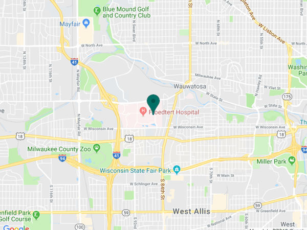
Eye Institute Google map location