Ophthalmology_Hero Image 2

Case Study 13 - CC: "Cannot see well" from left eye

all
Patient Visit

Patient History

HPI:
58 year-old administrative assistant Hispanic female complains of decreased vision in her left eye. She first noticed it when she closed her right eye while washing off her make-up about 2 weeks prior to her visit. The vision in her left eye is still blurred and dark and may have gotten slightly worse. She does have occasional symptoms of dry eyes with burning; for which she uses artificial tears. She denies any pain, redness, double vision, flashing lights, or new floaters.

Past Ocular History:
Slight myopia, uses glasses. No history of eye surgeries, amblyopia, strabismus, or eye trauma.

Ocular Medications:
Artificial tears occasionally

Past Medical History:
Menopause at 51, history of pulmonary embolism 5 years ago following L foot surgery, hypertension on medication

Surgical History:
L foot surgery after trauma

Past Family Ocular History:
Negative for macular degeneration, glaucoma or blindness.

Social History:
Married, 2 children. Smokes ½ ppd x 15 years. No alcohol or other recreational drugs.

Medications:
Calcium supplement, Multivitamin, Hydrochlorothiazide, Lisinopril

Allergies:
None

ROS:
Denies recent illness or any new CNS, heart, lung, GI, skin or joint symptoms.

Ocular Exam

Visual Acuity (cc):
OD: 20/25
OS: 20/200

IOP (tonoapplantation):
OD: 21 mmHg
OS: 21 mmHg

Pupils:
Equal, round and reactive to light, trace APD OS

Extraocular Movements:
Full OU. No nystagmus.

Confrontational Visual Fields:
Full to finger counting OD, inconsistent responses OS

External:
Normal both sides

Slit Lamp:

Lids and Lashes Normal OU
Conjunctiva/Sclera Normal OU
Cornea Clear OU
Anterior Chamber Deep and quiet OU
Iris Normal OU, no NVI OU
Lens 1+ NS OU
Anterior Vitreous Clear OU
Dilated Fundus Examination:
OD Clear view, CDR 0.5, with sharp optic disc margins; flat macula with normal foveal light reflex; slightly dilated retinal veins, mild arterial/venous nicking, normal peripheral retina
OS Clear view, edematous optic nerve with blurred margins; scattered intraretinal hemorrhages in all 4 quadrants, few cotton wool spots, macula thickening with loss of foveal light reflex, tortuous vessels.
Other:
OCT of the L eye - intra and subretinal fluid in the retina, distortion of the normal foveal architecture
Diagnosis and Discussion

Diagnosis
Central retinal vein occlusion (CRVO) L eye

Discussion

Differential Diagnosis:
This patient has suffered a central retinal vein occlusion (CRVO). The symptoms of sudden painless vision loss and the “blood and thunder” appearance of the retina (as seen in the picture) is typical of this diagnosis. Other diseases to consider include diabetic retinopathy, central retinal artery occlusion, retinopathy of anemia or leukemia or traumatic retinopathy.

Definition:
CRVO is the result of a blockage of the central retinal vein; most likely at the level of the lamina cribrosa. Typical retinal findings include diffuse intraretinal hemorrhages, retinal edema, and dilated tortuous retinal veins. Cotton wool spots, optic nerve edema and hemorrhages, and the subsequent development of neovascularization of the iris, optic nerve or retina can also be seen. CRVO can be classified into two broad categories: perfused (the most common type) or ischemic/non-perfused. Generally, perfused CRVO is a milder form of the disease, presenting with a better visual acuity and resulting in better final visual acuity and a decreased likelihood of iris or retinal neovascularization than ischemic/non-perfused CRVO. However, up to 1/3 of perfused CRVO may progress to the ischemic form. Some patients present with an intermediate form difficult to categorize. Vision loss due to CRVO can be due to macular edema, macular ischemia or complications from neovascularization including neovascular glaucoma or vitreous hemorrhage.

Examination:
Risk factors for CRVO include systemic hypertension, diabetes mellitus, vasculopathy and primary open angle glaucoma. A typical patient is male and 50 years or older. A comprehensive medical history is necessary to identify any risk factors and aid in referring to the appropriate medical care provider. Younger patients should also be evaluated for hypercoagulable states, other vascular diseases and the use of oral contraceptives, in addition to the above. An initial ophthalmic examination may include a fluorescein angiogram to determine the degree of ischemia/perfusion – although interpretation is hindered by the amount of retinal hemorrhages. Monthly examinations to include visual acuity, pupil check for APD and examination of the anterior segment angle for neovascularization for the first 3-6 months after diagnosis are necessary to identify cases of ischemic CRVO and of neovascularization that might require treatment.

Treatment:
Patients developing neovascularization need panretinal photocoagulation to prevent further complications. Macular edema associated with CRVO does not respond well to focal grid laser treatment, with visual acuity outcomes similar to those receiving no treatment. Treatment with intravitreal steroids or an anti-VEGF medication (ex. Avastin) have shown promising results.

The prognosis varies depending on whether the CRVO was ischemic or non-ischemic. Fifty percent of patients with non-ischemic CRVO will have vision better than 20/200 compared to only 10% of ischemic CRVO patients. A major complication of non-perfused CRVO is neovascularization, which occurs in approximately 60% of patients. Risk of other eye involvement is approx. 5%.

Self-Assessment Questions
  1. What are the typical symptoms in a patient suspected of having a central retinal vein occlusion (CRVO)?
  2. List the most common risk factors for the development of CRVO.
  3. In patients with CRVO which one would NOT be a cause of the decreased vision?

References/Resources

Self-Assessment Answers

1. What are the typical symptoms in a patient suspected of having a central retinal vein occlusion (CRVO)?
a. Hazy vision in one eye, floaters
Retinal pathology usually does not affect the anterior portion of the eye immediately.

2. List the most common risk factors for the development of CRVO.
b. Hypertension
Hypertension has been found to be a significant risk factor in the development of CRVO.

3. In patients with CRVO which one would NOT be a cause of the decreased vision?
d. Ptosis


Contact Ophthalmology

For patient care inquires, call us at (414) 955-2020 or use MyChart. Email is for research and education inquiries only.

Eye Institute Location

925 N. 87th St.

Milwaukee, WI 53226


Appointments

(414) 955-2020

(414) 955-6166 (fax)


Continuing Medical Education

Taylor Schmidt

taschmidt@mcw.edu

(414) 955-2088


Medical Education Coordinator

Ophth-Residency@mcw.edu


Associate Director of Development - Ophthalmology

Sarah Walker

sarawalker@mcw.edu

Refer to Us - Consultation requests

Patient Referral Form (PDF)

Fax to (414) 955-0136


Emergent Requests

Within 48 hours call

(414) 955-2020


Research

Vesper Williams

vewilliams@mcw.edu

(414) 955-7862


Advanced Ocular Imaging Program

aoip@mcw.edu

(414) 955-2647


Eye Institute Executive Director (Administrator)

Shannon Dreier
sdreier@mcw.edu

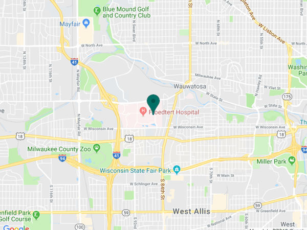
Eye Institute Google map location