Ophthalmology & Visual Sciences at the Eye Institute

Case Study 19 - CC: Wandering Eye and Double Vision

Original Author: Rebecca Mastey

all
Patient Visit

HPI:
8 year old female brought in by mother for evaluation of wandering eye and double vision. She was noted to have eye misalignment at age 1 and age 2 but exams were limited at that time. The patient had not followed with ophthalmology since then. Recently, the right eye has been wandering more frequently and has been noticed by teachers and students at school. She also gets double vision when she looks up and has adapted a slight chin up posture.

Past Ocular History:
Intermittent exotropia noted at age 1 and 2

Ocular Medications:
None

Past Medical History:
None

Surgical History:
None

Past Family Ocular History:
None

Social History:
Lives with parents and older sister. Normal growth and development for age.

Medications:
Allegra

Allergies:
NKDA, seasonal allergies

ROS:
Denied headache, denied recent illness


Ocular Exam

Visual Acuity (cc):
OD: 20/50 +1
OS: 20/50 +3

Pupils:

OD: Dark 8, Light 6, Round, Risk, No APD
OS: Dark 8, Light 6, Round, Risk, No APD

Extraocular Movements:
OD:  -3 to supraduction, otherwise full
OS: Full

Confrontational Visual Fields:
OD: Full
OS: Full

Slit Lamp Exam:

RIGHT EYE LEFT EYE
External Normal

Normal

Lids and Lashes Normal Normal
Conjunctiva/Sclera White and quiet White and quiet
Cornea Clear

Clear

Anterior Chamber Deep and quiet Deep and quiet
Iris Normal Normal
Lens Clear Clear
Anterior Vitreous Normal Normal
Dilated Fundus Examination:
RIGHT EYE LEFT EYE
Disc Normal Normal
C/D Ratio 0.2 0.2
Macula Normal Normal
Vessels Normal Normal
Periphery    

Imaging/additional tests:
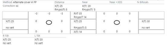
Sensorimotor exam:

  • Stereo: Animals 3/3, Circles 8/9
  • Abnormal head position (AHP): 5-10 degree chin up
  • Ophthalmic Case Study  Imaging Test
Diagnosis and Discussion

Diagnosis:
Monocular elevation deficiency

Differential Diagnoses:
The patient has monocular elevation deficiency (MED) in which there is a deficiency of one eye to elevate in all directions. It is likely congenital although not diagnosed until this visit due to limited exams on prior visits due to age. Other diagnoses on the differential include Brown syndrome, Duane syndrome, acquired elevation deficiency, Superior division of CN3 palsy, and congenital fibrosis of EOM. The patient history, as well as the sensorimotor exam and ductions help better understand the pattern and assist in narrowing the diagnosis to MED as the elevation deficit is the same in both abduction and adduction.

Definition:
Moncular elevation deficiency (MED) is defined as a limitation of elevation in one eye with normal movements in all other gazes. The superior limitation must be similar in both abduction and adduction. MED is relatively rare. The incidence of strabismus overall is about 5% and of these patients the incidence of MED is 0.5%. It is most commonly congenital although can be acquired. MED was formerly called “double elevator palsy” referring to the two ipsilateral elevator muscles (inferior oblique and superior rectus), however, this is becoming less widely used and replaced with MED. The pathophysiology is still poorly understood but leading theories include: superior rectus paresis, inferior rectus restriction, and/or unilateral supranuclear abnormality.

Examination:
MED often presents with vertical misalignment of the eyes which can cause an abnormal head posture (AHP), binocular diplopia, or amblyopia. Patients often adapt an AHP to offset diplopia that they notice. This adaptation helps the patient use both eyes together and therefore can prevent amblyopia. MED is a clinical diagnosis that is most notable on ductions and sensorimotor exam. There will be a limitation of elevation in the affected eye that is similar in all areas of upgaze. There can be ptosis and amblyopia in the affected eye although this is not required for the diagnosis. CT/MRI can rule out other etiologies since imaging is usually normal in MED.

Treatment:
As with any misalignment of the eyes, any underlying refractive error should be corrected with glasses and if there is amblyopia this should be addressed as well. This patient does not have diplopia or significant misalignment in primary gaze and therefore monitoring is sufficient for now. Close monitoring for any change in these symptoms (ie worsening diplopia, worsening alignment in primary, worsening head position, or loss of stereovision) would warrant surgical intervention. Surgically, the goal would be to improve the position of the affected eye in primary gaze and increase the field of binocular vision.

Self-Assessment Questions
How do you differentiate between MED and Brown Syndrome?
a. MED has elevation deficit in adduction only and Brown syndrome has elevation deficit in adduction and abduction
b. Brown Syndrome has elevation deficit in adduction only and MED has elevation deficit in adduction and abduction
c. MED has elevation deficit in adduction only and Brown syndrome has depression deficit in adduction only
d. MED has elevation deficit in one eye and Brown Syndrome has elevation deficit in both eyes

True or False: Patients with MED who adapt a head up posture is a sign of amblyopia

Which is a suggested cause of MED?
a. Superior rectus paresis
b. Inferior rectus restriction
c. Unilateral supranuclear abnormality
d. All the above
Self-Assessment Answers

How do you differentiate between MED and Brown Syndrome?
b. Brown Syndrome has elevation deficit in adduction and MED has elevation deficit in adduction and abduction
Correct answer is B. MED has elevation deficit that is comparable in all fields of upgaze (abduction and adduction) whereas Brown syndrome has an elevation deficit only in adduction.

True or False: Patients with MED who adapt a head up posture is a sign of amblyopia
This statement is false. Patients who adapt a head up posture are doing so in order to use both eyes together. This is actually a reassuring sign of fusion, preserving stereovision, and avoiding amblyopia. If the affected eye is amblyopic, the patient may not adapt a head up posture as they are not using the eyes together.

Which is a suggested cause of MED?
d. All the above
Correct answer is D. The pathophysiology is still poorly understood and all of the above causes could be the underlying reason for MED in a given patient. If the patient warrants surgical intervention, it is especially important to try and further evaluate which of these three is the cause in order to perform the correct surgerical intervention.

References/Resources:

  • Color Atlas of Strabismus Surgery: Strategies and Techniques, Third Edition, Kenneth W Wright
  • Gorey, Dhiman, Thacker, et al (2015). Monocular Elevation Deficit-Simplified. Delhi Journal Of Ophthalmology. 26. 7-13. 10.7869/djo.127.
  • https://eyewiki.org/Monocular_Elevation_Deficit